Nuclear envelope Biology Diagrams An in vitro nuclear disassembly system reveals a role for the RanGTPase system and microtubule-dependent steps in nuclear envelope breakdown. J. Cell Biol. 178 , 595-610 (2007). The COPI complex functions in nuclear envelope breakdown and is recruited by the nucleoporin Nup153. Dev Cell. 2003;5:487-498. doi: 10.1016/s1534-5807(03)00262-4. This paper implicates the Golgi COPI vesiculation machinery in breakdown of the nuclear envelope. In addition, COPI recruitment to the nuclear envelope is facilitated by its
The mechanism of nuclear envelope breakdown (NEBD) was investigated in live cells. Early spindle microtubules caused folds and invaginations in the NE up to one hour prior to NEBD, creating mechanical tension in the nuclear lamina. The first gap in the NE appeared before lamin B depolymerization, at the site of maximal tension, by a tearing mechanism.
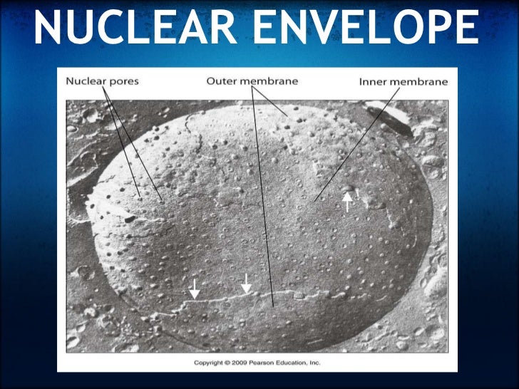
Nuclear envelope: Current Biology Biology Diagrams
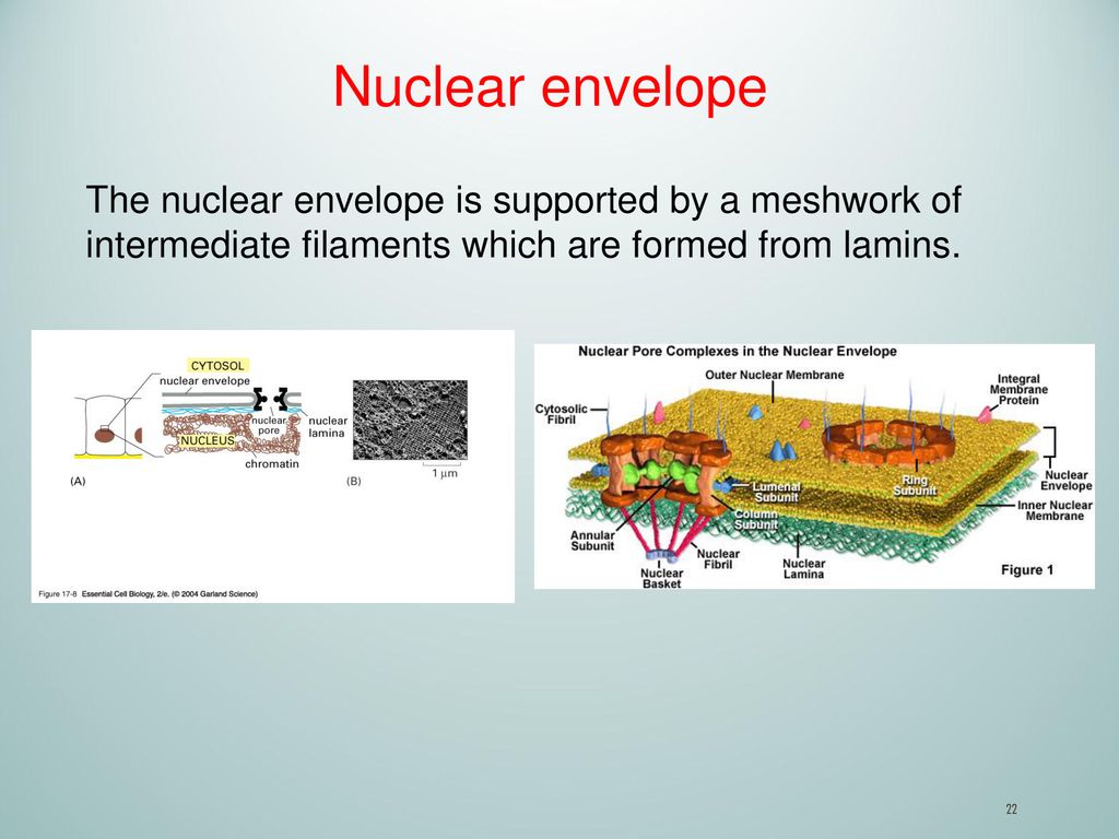
Breakdown and reformation of the nuclear envelope (NE) during cell division is one of the most dramatic structural and functional changes in higher eukaryotic cells. NE breakdown (NEBD) marks a highly regulated switch in chromosome confinement by membranes in interphase to microtubules in M-phase. The boundary of interphase nuclei has a rigid and highly interconnected architecture made up of a Nuclear envelope breakdown in starfish oocytes proceeds by partial NPC disassembly followed by a rapidly spreading fenestration of nuclear membranes. J Cell Biol 160:1055-1068 [PMC free article] [Google Scholar] Liu J, Prunuske AJ, Fager AM, Ullman KS 2003. The COPI complex functions in nuclear envelope breakdown and is recruited by the

Through this process, the nuclear envelope projects tubules that capture damaged DNA, mediating its repair. Current models suggest that DNA double-strand breaks (DSBs) can move to the nuclear

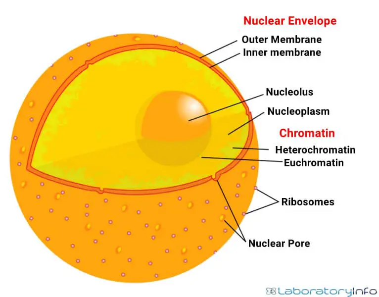
The Nuclear Envelope Biology Diagrams
Nuclear envelope breakdown occurs by stepwise disassembly of nuclear pore complexes, inner nuclear membrane proteins and, finally, lamins, and is thought to be driven mostly by the phosphorylation of these proteins by mitotic kinases. In somatic cells, nuclear envelope breakdown is additionally facilitated by mitotic microtubules, which pull on An in vitro nuclear disassembly system reveals a role for the RanGTPase system and microtubule-dependent steps in nuclear envelope breakdown. J. Cell Biol. 178 , 595-610 (2007).