Meniscus Tear Austin TX Biology Diagrams The anterior cruciate ligament (ACL), is the weaker of two cruciate ligaments of the knee, the other being the posterior cruciate ligament.These intracapsular ligaments are so named due to the fact that they cross each other, creating an imaginary cross (the word cruciate comes from the latin word crux that means cross). Structure and Function of the Knee Meniscus 2.1 Meniscus Anatomy. The knee joint contains the meniscus structure, Transepiphyseal replacement of the anterior cruciate ligament in skeletally immature patients. A preliminary report. J Bone Jt Surg Am. 2003;85-A:1255-63. doi: 10.2106/00004623-200307000-00011. The anterior cruciate ligament (ACL) is one of 2 cruciate ligaments that aids in stabilizing the knee joint. It is a strong band made of connective tissue and collagenous fibers that originate from the anteromedial aspect of the intercondylar region of the tibial plateau and extends posterolaterally to attach to the medial aspect of the lateral femoral condyle, where there are two important
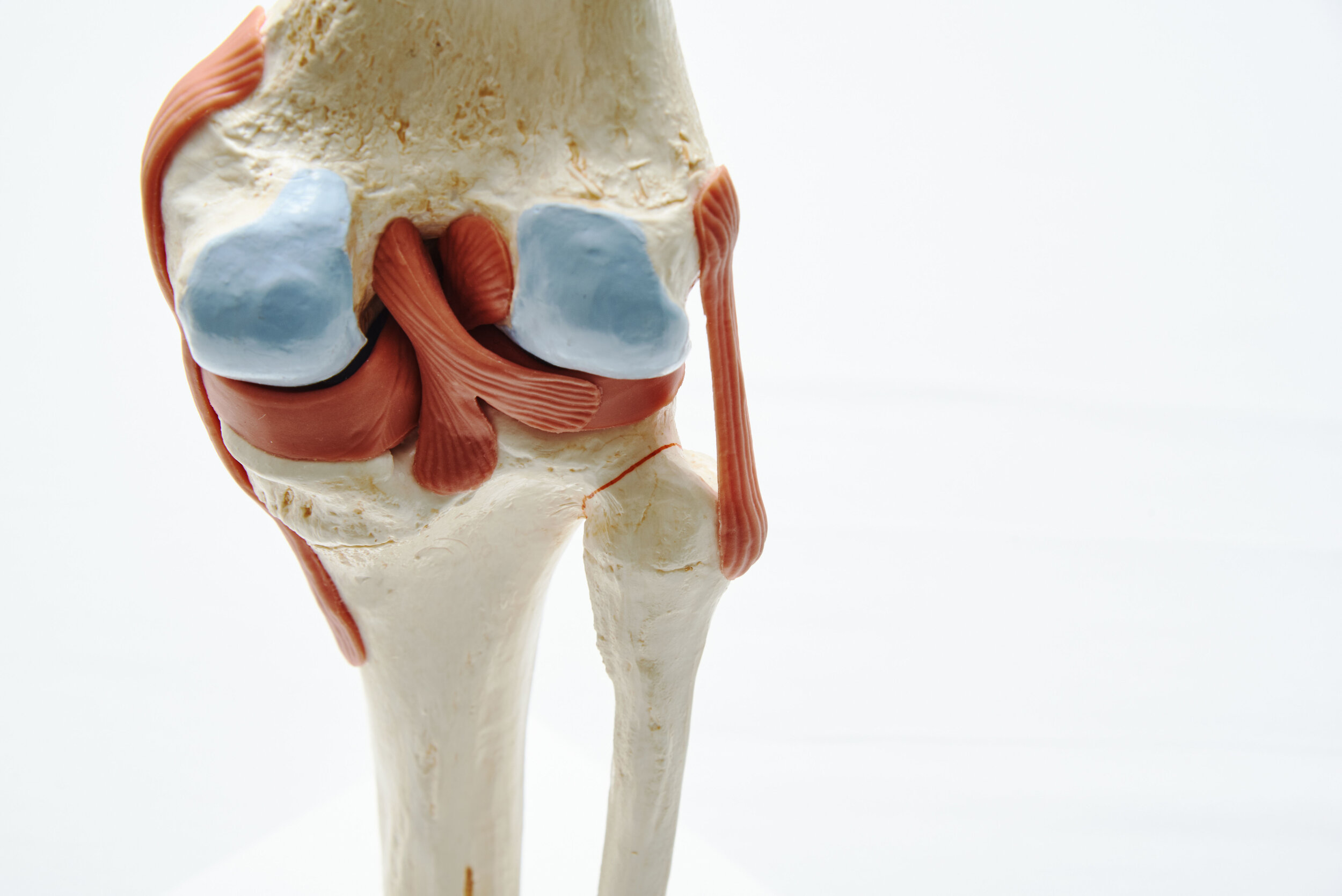
Medial Meniscus: C-shaped and larger, it attaches firmly to the medial collateral ligament (MCL).; Lateral Meniscus: O-shaped and more mobile. [4] Functionally, the menisci act as shock absorbers, enhance joint stability, and improve congruence between the femur and tibia.. Ligaments. The knee joint is stabilized by several strong ligaments, categorized into extracapsular and intracapsular Information about the anatomy of the knee which connects the shinbones to the femur with the knee cap (patella), quadricpes muscles, hamstring muscles, anterior cruciate ligament (ACL), posterior cruciate ligament (PCL), medial collateral ligament(MCL) and lateral collateral ligament (LCL), lateral meniscus, medial meniscus ACL Knee Anatomy. Anatomy. The anterior cruciate ligament (ACL) is 1 of 4 main ligaments in the knee. Ligaments are rope-like structures that connect and hold the bones together to keep the knee stable. The ACL, along with the posterior cruciate ligament (PCL), is located in the center of the knee. (ACL) and her meniscus, wrestler Sierra
Anterior cruciate ligament: Anatomy and function Biology Diagrams
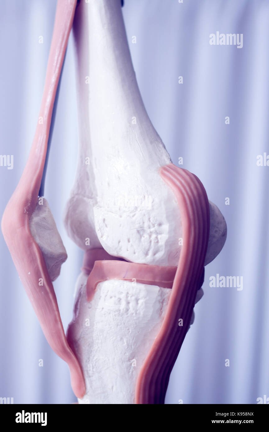
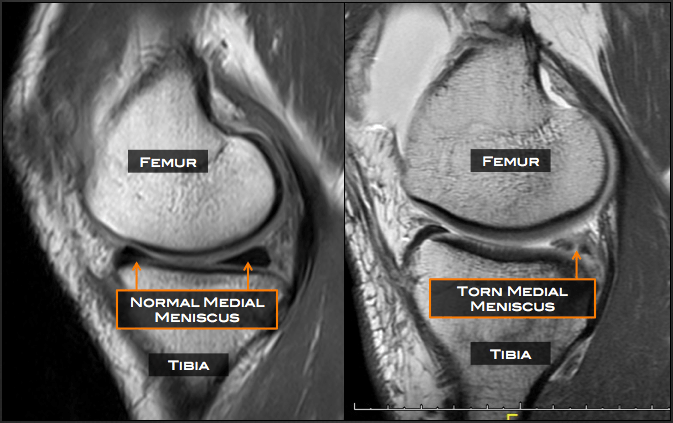
The knee meniscus is a special layer of cartilage that lines the knee joint. The job of the meniscus is to cushion the knee joint and transfer forces between the tibia and femur, the thigh and shin bones. Most of the joints in our body are lined with a thin layer of articular cartilage, made of collagen and chondroitin. This provides a smooth
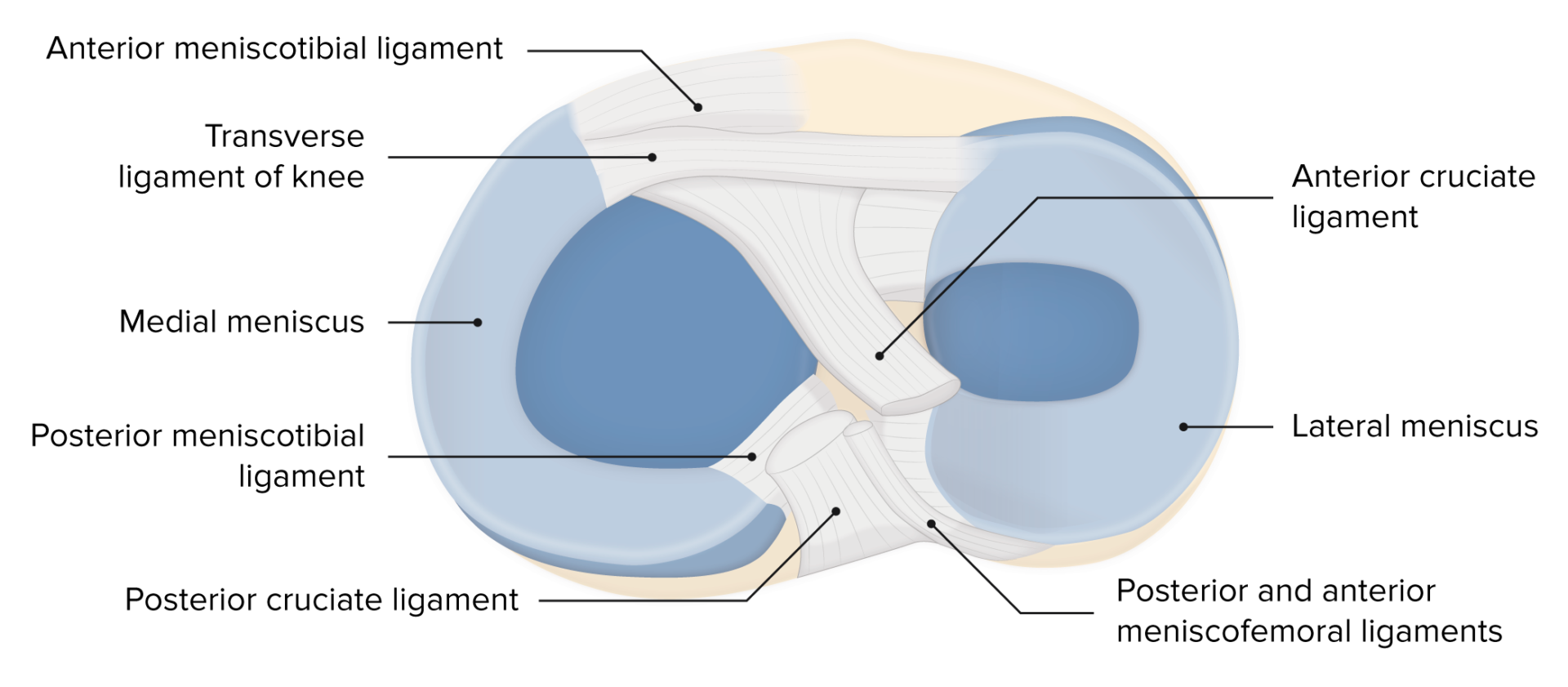
The anterior cruciate ligament (ACL) is one of the two cruciate ligaments which stabilizes the knee joint by preventing excessive forward movements of the tibia or limiting rotational knee movements. It is one of the most commonly injured structures in sports medicine, and yet it, unfortunately, does not heal when damaged.[1] This article presents the anatomy and function of the ACL to help
Anterior Cruciate Ligament Knee Injury Biology Diagrams
Information about the anatomy of the knee which connects the shinbones to the femur with the knee cap (patella), quadricpes muscles, hamstring muscles, anterior cruciate ligament (ACL), posterior cruciate ligament (PCL), medial collateral ligament(MCL) and lateral collateral ligament (LCL), lateral meniscus, medial meniscus