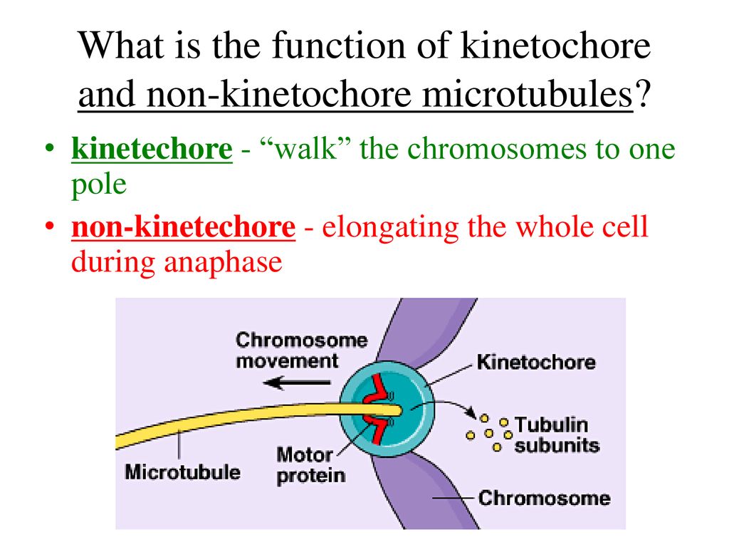
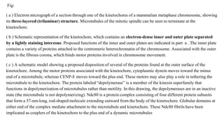
Kinetochore Biology Diagrams 1. An Overview of Kinetochore Structure and Functions In eukaryotes, the kinetochore is a proteinaceous multi-subunit assembly whose main function is to generate load-bearing attachments of sister chromatids (the replicated chromosomes held together by the protein complex cohesin) to spindle microtubules during cell division (mitosis or meiosis) (Figure 1 A). Kinetochores couple sister 381 Kinetochore function: molecular motors, switches and gates Tim J Yen* and Bruce T Schaart Kinetochores are essential for accurate chromosome grow and shorten in a coordinated fashion to move the segregation. Recent studies reveal that vertebrate chromosome throughout mitosis (see Fig. 1 and [6-8]). kinetochores are sophisticated propulsion systems composed This bidirectional movement of

Kinetochores are dynamic complexes containing MT motor and cell-cycle regulatory proteins, which serve three functions during cell division. They attach each replicated chromosome to the opposing poles of the mitotic spindle, help position the chromosome on the spindle and then inhibit chromatid separation (and anaphase onset) until all of the Kinetochore proteins can be grouped according to their concentration at kinetochores during mitosis: some proteins remain bound throughout cell division, whereas some others change in concentration. Furthermore, they can be recycled in their binding site on kinetochores either slowly (they are rather stable) or rapidly (dynamic).
lncreased risk of slippage upon disengagement of the mitotic checkpoint Biology Diagrams
During mitosis, it's up to the kinetochores assembled on the centromeres of each chromosome to give a cell the go-ahead to begin anaphase. A kinetochore will only give its ready signal once it becomes attached to and stretched apart by microtubules emanating from both opposing spindle poles. This process, known as the spindle checkpoint, ensures the even distribution of chromosomes between At the onset of mitosis, rapidly growing and shrinking microtubules (MTs) probe the cytoplasm in search of kinetochores, which are macromolecular complexes assembled on opposite sides of the centromere that mediate interactions between the chromosomes and MTs.

Introduction During mitosis, sister chromatids are segregated to the two daughter cells. This process requires the interaction between specialized chromosomal structures (kinetochores) and polymers of alpha- and beta-tubulin called microtubules. Abstract A critical requirement for mitosis is the distribution of genetic material to the two daughter cells. The central player in this process is the macromolecular kinetochore structure, which binds to both chromosomal DNA and spindle microtubule polymers to direct chromosome alignment and segregation. This review will discuss the key kinetochore activities required for mitotic chromosome In mitosis, chromosomes condense and the two copies become visible as "sister chromatids". One kinetochore is assembled on each of the two sister chromatids of a chromosome, and both sister kinetochores become attached to opposite spindle poles by metaphase.