
Esophagus anatomy Muscular tube that carries food and liquid from the Biology Diagrams The Epithelium: The inside of the esophagus is made up of cells called stratified squamous epithelium.; The Lamina Propria: This is a layer of connective tissue just under the epithelium.This layer contains lymphocytes. Cells that are an important part of our immune system. Muscularis Mucosa: This is a layer of smooth muscle that is essential in helping nutrients flow into the submucosa.
Treatment for esophagus problems depends on the cause. Some esophagus problems can be treated with over-the-counter medication or diet changes. Other conditions may require prescription medication, procedures or surgery. Common medications for esophagus conditions include: Antacids: Antacids neutralize stomach acids. Some common brand names Its main functions include: Transporting food: The esophagus moves food from the mouth to the stomach through a process called peristalsis, where coordinated contractions of the esophageal muscles push the food downward. Protection against reflux: The lower esophageal sphincter is a ring of muscle located at the bottom of the esophagus that helps to prevent stomach acid and food from flowing Anatomy of the Esophagus. The esophagus is a muscular tube about ten inches (25 cm.) long, extending from the hypopharynx to the stomach.The esophagus lies posterior to the trachea and the heart and passes through the mediastinum and the hiatus, an opening in the diaphragm, in its descent from the thoracic to the abdominal cavity.The esophagus has no serosal layer; tissue around the esophagus

The Esophagus: Anatomy and 3D Illustrations Biology Diagrams
This document provides an overview of the development, anatomy, and physiology of the esophagus. It discusses: 1. The embryological development of the pharynx, esophagus, and trachea from the buccopharyngeal membrane and branchial arches. 2. The anatomy of the esophagus including its course, layers, blood supply, nerve supply, and sphincters. 3.

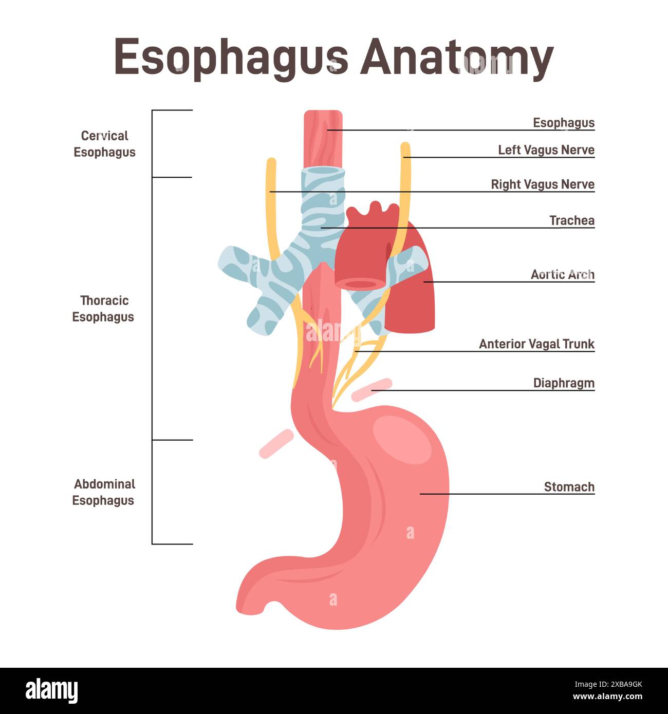
The oesophagus is a fibromuscular tube, approximately 25cm in length, that transports food from the pharynx to the stomach.. It originates at the inferior border of the cricoid cartilage (C6) and extends to the cardiac orifice of the stomach (T11).. In this article we shall examine the anatomy of the oesophagus - its structure, vascular supply and clinical correlations. Esophagus (anterior view) The esophagus (oesophagus) is a 25 cm long fibromuscular tube extending from the pharynx (C6 level) to the stomach (T11 level). It consists of muscles that run both longitudinally and circularly, entering into the abdominal cavity via the right crus of the diaphragm at the level of the tenth thoracic vertebrae.. It actively facilitates the passage of the food bolus The esophagus, historically also spelled oesophagus, is a tubular, elongated organ of the digestive system which connects the pharynx to the stomach. The esophagus is the organ that food travels through to reach the stomach for further digestion. It follows a path that travels behind the trachea and heart, in front of the spinal column, and through the diaphragm before entering the stomach.[1][2]

Anatomy of the Esophagus Biology Diagrams
Anatomy of Oesophagus The oesophagus is a fibromuscular tube approximately 25 cm long in adults, extending from the lower end of the pharynx (C6) to the stomach's cardiac end (T11). It runs vertically downwards with slight leftward inclinations at the thoracic inlet and near the diaphragm (T7-T10).