
Anatomy Of Carotid Artery Biology Diagrams The carotid arteries extend out from the aorta artery, which transports blood out of the heart and is the body's largest artery. The carotid arteries carry blood through the neck up to the brain. Stenosis means "narrowing." Narrowed carotid arteries usually occur due to atherosclerosis. This is an accumulation of plaque along your artery walls. In your carotid arteries, this plaque often forms near the carotid bifurcation. Plaque buildup raises your risk of a transient ischemic attack (TIA) or a stroke. That's because blood clots The carotid body is a 2 to 6 mm, round bilateral sensory organ in the peripheral nervous system located in the adventitia of the bifurcation of the common carotid artery. These peripheral chemosensory cells play a vital, physiologic role in the human body as they maintain physiologic homeostasis and regulation of sustaining life. These sensory chemoreceptors act to detect chemical changes in

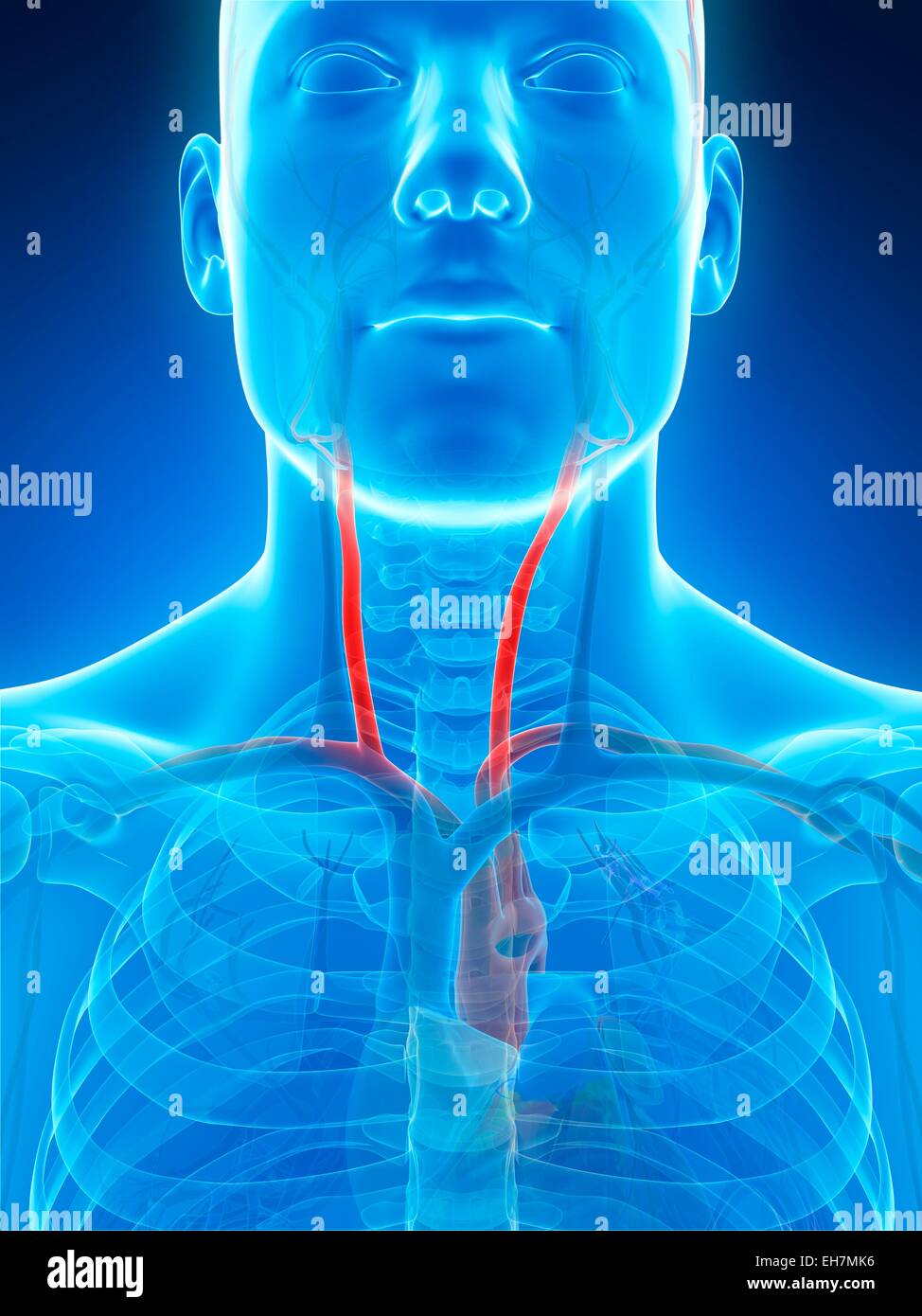
The carotid arteries are the primary vessels supplying blood to the brain and face.[1][2] The right common carotid artery (RCCA) originates in the neck from the brachiocephalic artery while the left common carotid artery (LCCA) arises in the thorax from the arch of the aorta.[3] The carotid body chemoreceptor is sensitive to decreased PO2 The arteries are essential for carrying blood enriched with oxygen from the heart to other regions of the body. Anatomy of Common Carotid Artery. The body's main artery, the aorta, from which the carotid artery extends, is responsible for carrying out blood from the heart.
5 Largest Arteries in the Human Body Biology Diagrams
The carotid body is situated on the posterior aspect of the bifurcation of the common carotid artery. [3]The carotid body is made up of two types of cells, called glomus cells: glomus type I cells are peripheral chemoreceptors, and glomus type II cells are sustentacular supportive cells.. Glomus type I cells are derived from the neural crest. [4] They release a variety of neurotransmitters
Some of the most important conduits of blood flow are the carotid arteries, which consist of the common carotids, external carotids, and the internal carotids. Just like all arteries in the human body, the internal carotids consist of three layers histologically. Going from inside to outside the vessel, they include the tunica intima Common Carotid Artery; Diameter: 4.3 mm-7.7 mm Blood flow: 350-550 milliliters per minute Common problem: Cholesterol plaques Location: Neck and head. Source: wikimedia.org. The aorta is the largest artery in the human body, as well as the main artery in the circulatory system.

Anatomy, Course, Branches, Function Biology Diagrams
Carotid artery, one of several arteries that supply blood to the head and neck. Of the two common carotid arteries, which extend headward on each side of the neck, the left originates in the arch of the aorta over the heart; the right originates in the brachiocephalic trunk, the largest branch from Facts You Should Know: The Human Body Quiz. The common carotid artery is a major branch of the carotid artery, one of the key blood vessels in the human body. It plays a critical role in supplying oxygenated blood to the head and neck regions. Let's explore the specific characteristics, functions, and potential health conditions associated with the common carotid artery.
