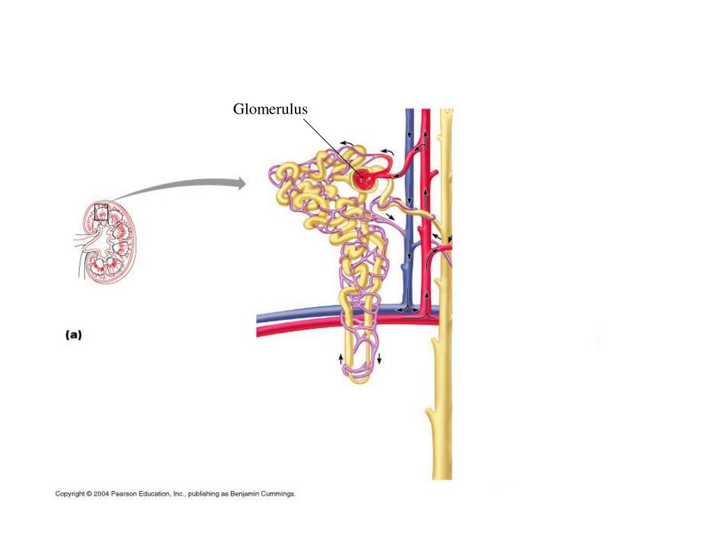
1 glomerular disease anatomy pysiology of kideny Biology Diagrams Describe the histology of the proximal convoluted tubule, loop of Henle, distal convoluted tubule, and collecting ducts capsule. The glomerulus is a high-pressure capillary bed between afferent and efferent arterioles. Bowman's capsule surrounds the glomerulus to form a lumen, and captures and directs this filtrate to the PCT
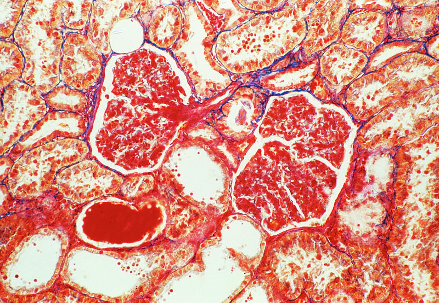
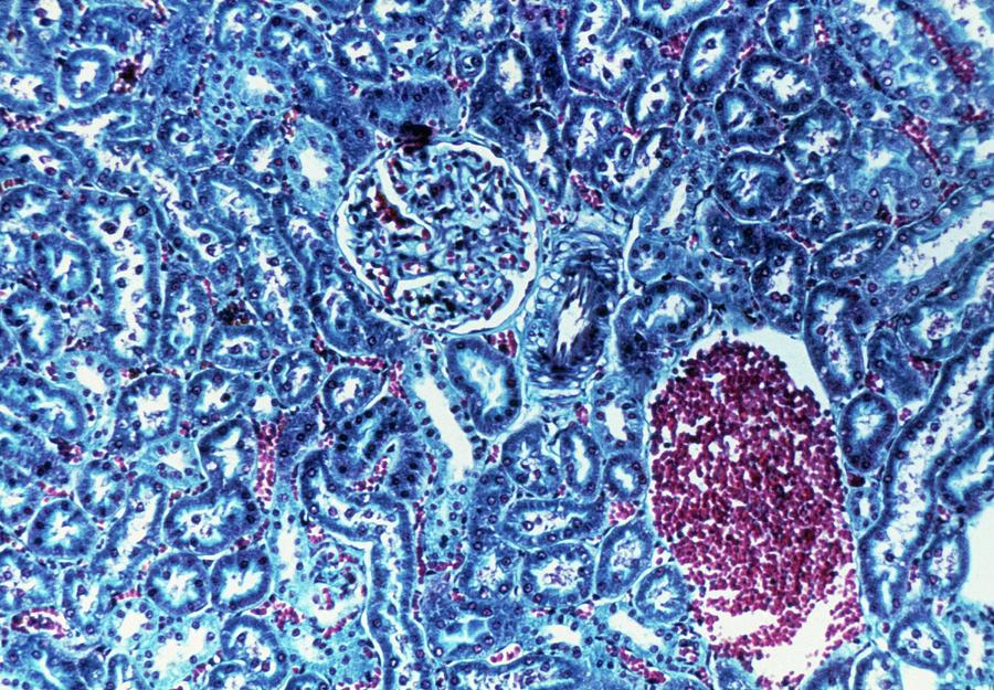
The renal corpuscle is the initial part of the nephron, located in the renal cortex, where blood filtration occurs.[2] It consists of a tuft of capillaries called the glomerulus and a surrounding cup-shaped structure known as Bowman's capsule. The renal corpuscle acts as a filtration unit, removing water, ions, and waste products from the blood The glomerulus (pl.: glomeruli) is a network of small blood vessels (capillaries) known as a tuft, located at the beginning of a nephron in the kidney.Each of the two kidneys contains about one million nephrons. The tuft is structurally supported by the mesangium (the space between the blood vessels), composed of intraglomerular mesangial cells.The blood is filtered across the capillary walls The glomerulus is the main filtering unit of the kidney. Learn everything about its anatomy and functions now on Kenhub! Connection lost. Please refresh the page. Online The ultrafiltrate is collected in the Bowman's space and drains directly into the proximal tubule of the nephron. The glomerulus is composed mainly of three cell types:
TeachMePhysiology Biology Diagrams
The first part of the collecting tubule (cortical collecting tubule) is within the renal cortex while the latter part (medullary collecting tubule) is within the renal medulla. Diagram of the Nephron, Glomerulus and Different Parts of the Tubule. The tubules are lined with a thin layer of epithelial cells. The afferent arteriole (at the proximal glomerulus) dilates, while the efferent arteriole (at the distal glomerulus) constricts. This creates a pressure gradient throughout the glomerulus, causing filtration under pressure. The filtration rate of molecules of the same charge across the filtration barrier is inversely related to their molecular
Anatomy. The kidney is a complex organ with a unique structure, made up of various internal and external components that work together to support its vital functions. which surrounds a network of capillaries called the glomerulus. Together, they form the renal corpuscle, where blood filtration occurs. Proximal Convoluted Tubule (PCT

Structure, Location, Function, Diagram, Anatomy Biology Diagrams
The kidneys have several essential homeostatic functions. These functions include waste removal (NH3), fluid/electrolyte balance, metabolic blood acid-base balance, as well as producing/modifying hormones for blood pressure, calcium/potassium homeostasis, and red blood cell production. The renal corpuscle (filtration unit, which comprises the glomerulus and the surrounding glomerular or Bowman The renal corpuscle is a small, round-shaped component of the nephron, located in the renal cortex of kidneys.Each renal corpuscle is made up of two structures: a small tuft of capillaries known as the glomerulus, and a surrounding cup-shaped structure known as the Bowman's capsule (glomerular capsule). In total, there are approximately one-million renal corpuscles in each kidney.